One Breast Cancer, Three Questions: A Walkthrough on Pan.bio
Three breast cancer analyses on public data: tumour vs normal variant calling, lab-tested BRCA1 variants and 1,066 TCGA tumours, run end-to-end on Pan.bio.

 Thikra Daraghmeh

On this pageTumour-normal variant calling: what was inherited, what the tumour acquired
A public breast cancer cell line called HCC1395 carries a BRCA1 variant that stops the gene short. The same variant sits in normal cells from the same woman, so it was there before the cancer. In the tumour, next to it, is one of the best-known TP53 changes in cancer. One pipeline run on Pan.bio’s workflows found both.
Breast cancer genomic analysis rarely stops at one question. What did this person inherit? Does this particular variant break the gene? How does one tumour compare with a thousand others? Each question usually lives in a different tool, a different environment, and often with a different team.
This walkthrough runs all three on public data, in one platform, and shows each step: Pan.bio Workflows for the raw sequencing reads, Pan.bio Notebooks with bioMind for the analysis, and VAIC for variant interpretation. No patient records are involved, and every number below comes from the run shown beside it.
Tumour-normal variant calling: what was inherited, what the tumour acquired
Two kinds of genetic test answer two different questions: a germline test reads normal cells, usually from blood, for variants a person was born with and can pass on, while a tumour test reads the cancer for changes that arose inside it. Sequencing tumour and normal cells from the same person and comparing them separates the two.
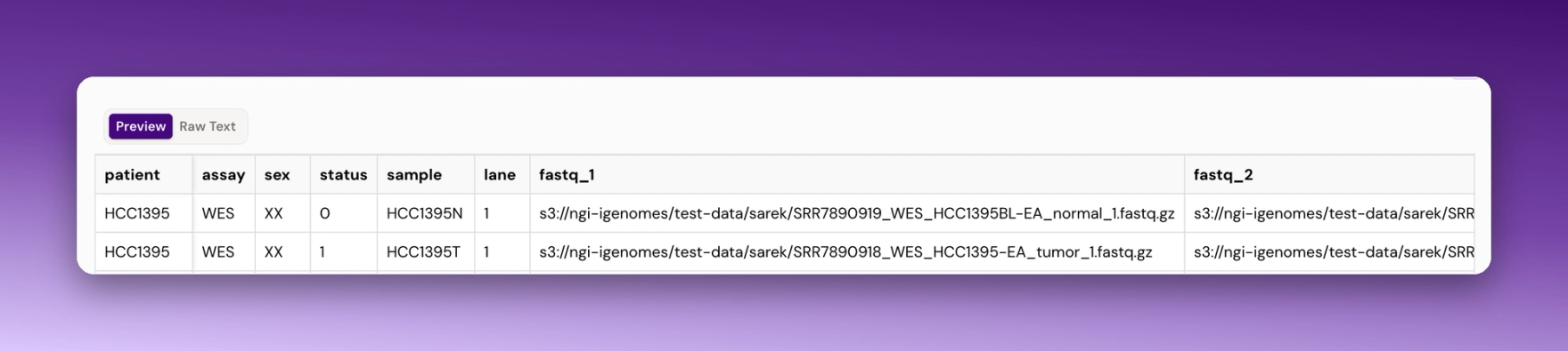
HCC1395 makes this possible on public data; it is a breast cancer cell line, and HCC1395BL is a normal cell line from the same woman. The SEQC2 consortium uses the pair as a community reference for testing how well pipelines find cancer mutations [3], we started from their whole-exome sequencing reads (SRR7890919 for the normal, SRR7890918 for the tumour), listed in a sample sheet with one row per sample: status 0 for normal, 1 for tumour (Figure 1).
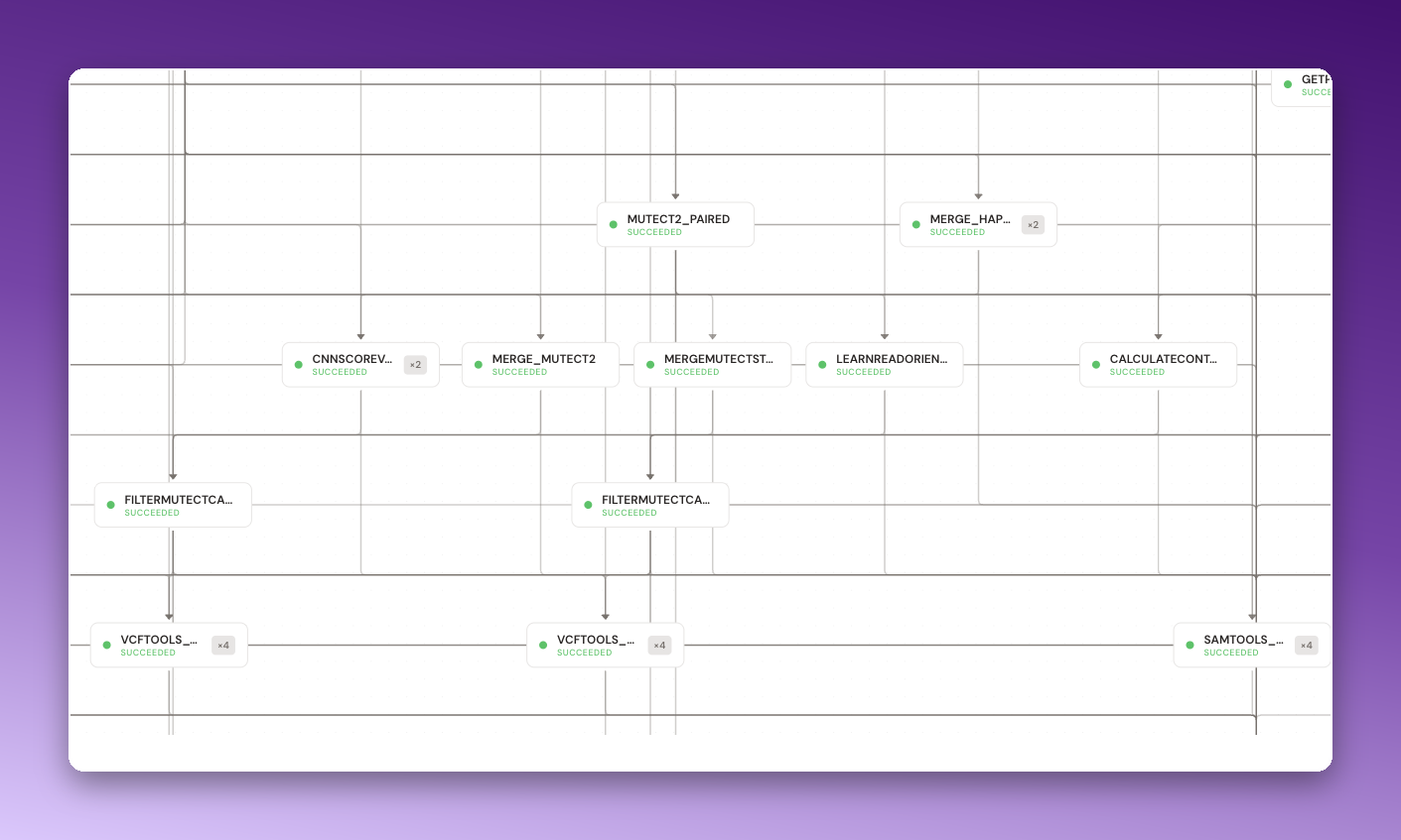
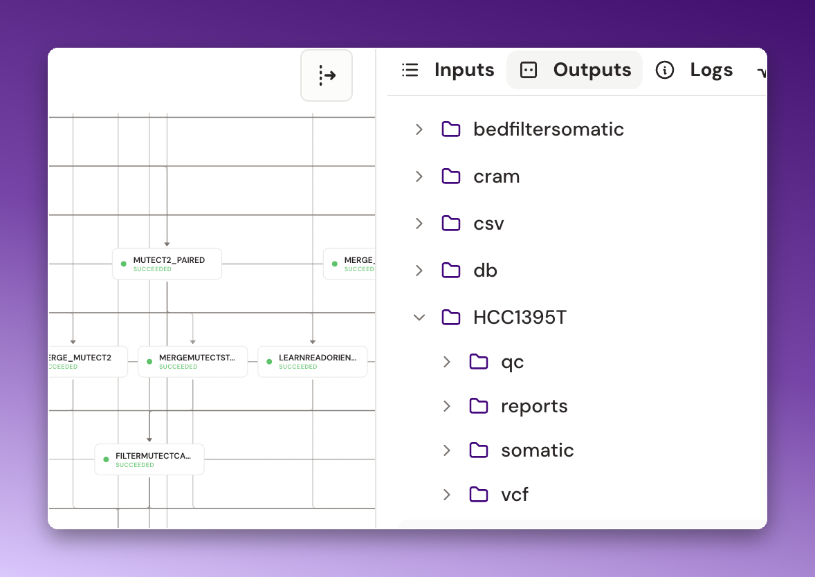
One run of a Pan.bio variant-calling workflow built on nf-core/sarek handled the rest (Figure 2). It checked read quality, aligned the reads to GRCh38, called inherited variants in the normal with GATK HaplotypeCaller, and called tumour variants against the normal with Mutect2, it then annotated every call against ClinVar, gnomAD v4.1, Ensembl VEP 115, REVEL and AlphaMissense, checked coverage across the ACMG secondary-findings genes, and wrote a report (Figure 3).
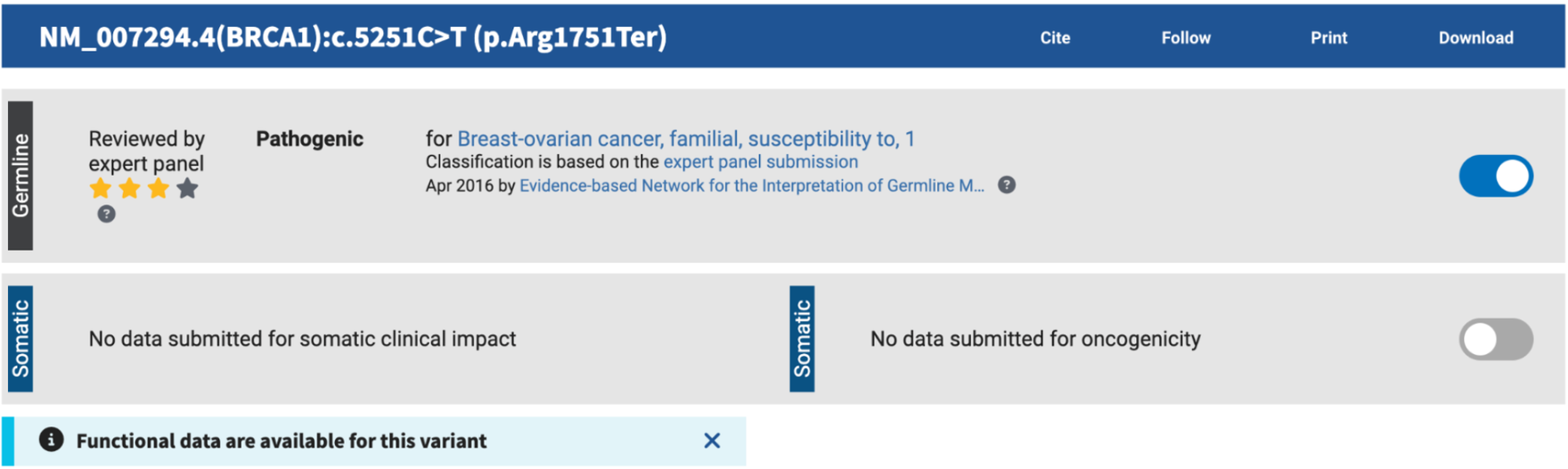
In the normal cells: BRCA1 c.5251C>T, p.(Arg1751Ter), on one of the two copies (93 of 202 reads). The change turns an arginine codon into a stop signal, so the protein is cut short before the second of its two BRCT repeats. ClinVar lists it as pathogenic, reviewed by an expert panel (Figure 4).
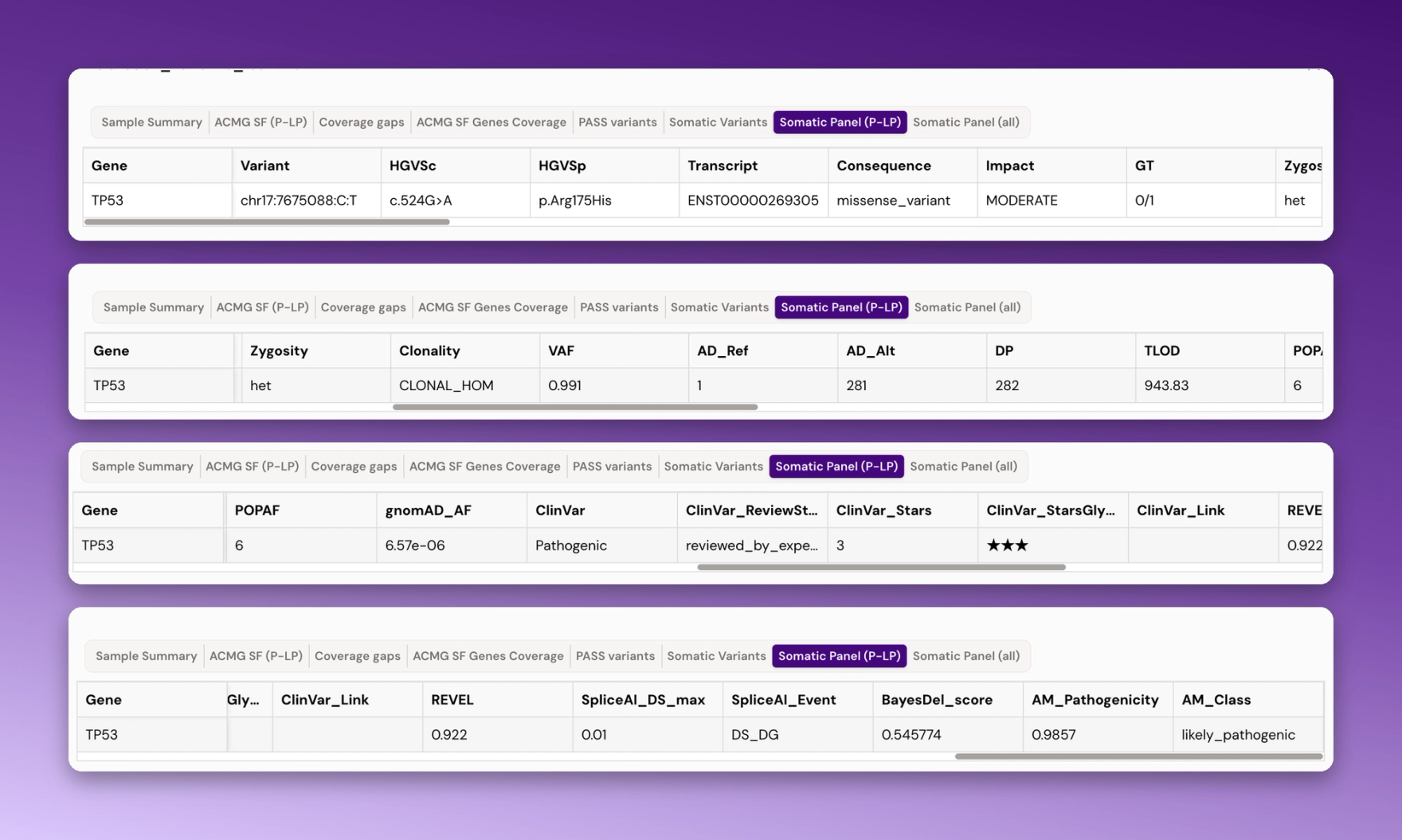
In the tumour: TP53 c.524G>A, p.(Arg175His), in 281 of 282 reads, so the tumour cells carry no normal copy at that position. ClinVar lists it as pathogenic, reviewed by an expert panel; REVEL scores it 0.922 and AlphaMissense 0.986 (Figure 5).
The run averaged 168.62x coverage across the targeted genes, with 100% of reads mapped. An earlier screen of six basal-like breast cancer cell lines described HCC1395 as BRCA1-deficient [5]. An inherited BRCA1 stop variant is consistent with that, although the earlier study does not name the variant.
A report that says “pathogenic in ClinVar” is a starting point. The next question is what evidence stands behind that word.
When the evidence comes from a lab, not a family tree
Classifying a variant means weighing several kinds of evidence: how common it is in the population, what other labs have reported, what prediction tools expect, and what happens to the protein when someone tests it [4]. The last kind is the hardest to come by. For most rare variants, nobody has measured anything.
For two regions of BRCA1, someone has. In 2018, Findlay and colleagues edited 3,893 single-letter changes into BRCA1 in human cells, covering the RING region (exons 2 to 5) and the BRCT region (exons 15 to 23), and measured which ones stopped the gene working [1]. Checked against variants already classified in ClinVar, the assay picked out 96.7% of the pathogenic ones at 98.2% specificity [1].
We asked bioMind, the AI agent inside Pan.bio Notebooks, to load that dataset and count the results by kind of change (Figure 6). It wrote the code; we reviewed it and ran it (Figure 7).
Table 1. BRCA1 variants tested by Findlay et al. [1], by kind of change.
Kind of change | Tested | Broke the gene | Share |
|---|---|---|---|
Missense (one amino acid swapped) | 2,086 | 441 | 21.1% |
Nonsense (early stop) | 138 | 136 | 98.6% |
Intronic or splice region | 1,034 | 235 | 22.7% |
Synonymous (“silent”) | 579 | 11 | 1.9% |
5′ UTR | 56 | 0 | 0% |
About one in five missense changes broke the gene (Table 1). So did 11 synonymous changes, which leave the protein sequence untouched; the likeliest explanation is that they disturb how the gene's RNA is spliced.
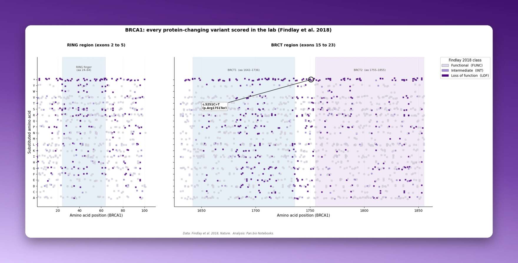
bioMind then drew every protein-changing variant as a map (Figure 8). Position along the protein runs left to right, the new amino acid runs up the side, and the top row (*) holds changes that create a stop signal. Dark purple squares broke the gene; pale ones left it working. The top row is almost solid dark, matching the 136 of 138 stop changes in the table. One square is circled: c.5251C>T, the inherited variant from use case 1, which the experiment scored as broken (−1.88).
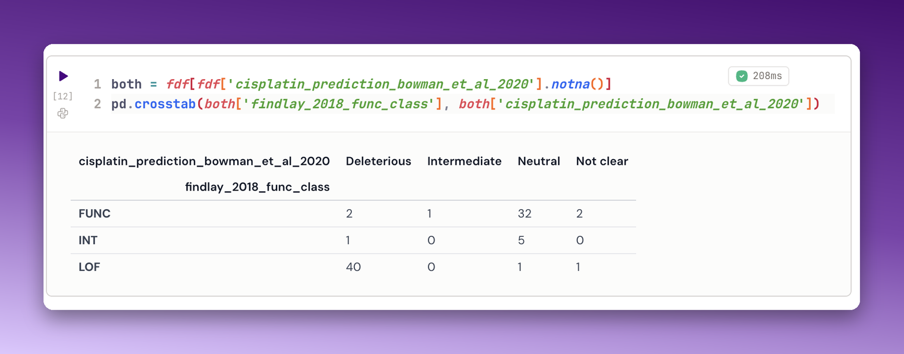
A second lab tested 238 BRCA1 variants for their ability to restore DNA repair, using cisplatin, olaparib and DR-GFP assays [2]. Of the 42 variants that Findlay scored as broken and that this lab also tested, 40 were deleterious in the cisplatin assay, and of the 37 Findlay scored as working, 32 came back neutral (Figure 9). Two methods, two labs, largely the same answer.
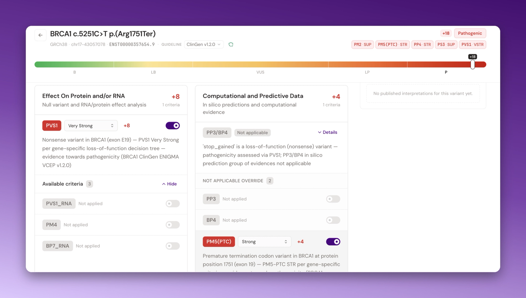
From lab data to a classification. VAIC applies the rules the ClinGen ENIGMA expert panel wrote for BRCA1 and BRCA2 (v1.2.0) and shows which criterion each piece of evidence supports. We searched three variants (Table 2).
Table 2. Three BRCA1 variants classified in VAIC, with their ClinVar expert panel classification.
Variant | VAIC result | Lab evidence, as VAIC weighs it | ClinVar |
|---|---|---|---|
c.5251C>T p.(Arg1751Ter), the inherited variant | Pathogenic (+18) | PS3, supporting | Pathogenic, expert panel |
c.181T>G p.(Cys61Gly), RING region | Pathogenic (+14) | PS3, strong | Pathogenic, expert panel |
c.4956G>A p.(Met1652Ile), BRCT region | Benign (BA1) | BS3, strong | Benign, expert panel |
The three rows show three different roles for the same kind of evidence:
- c.5251C>T: the stop signal does most of the work (PVS1, very strong). The lab result adds a supporting point on top (Figure 10).
- c.181T>G: a missense change with no stop signal, so the lab result counts as strong evidence. Even with a supporting point for benign from its frequency in gnomAD, VAIC classifies it as pathogenic, matching the expert panel’s ClinVar entry (Figures 11 and 12).
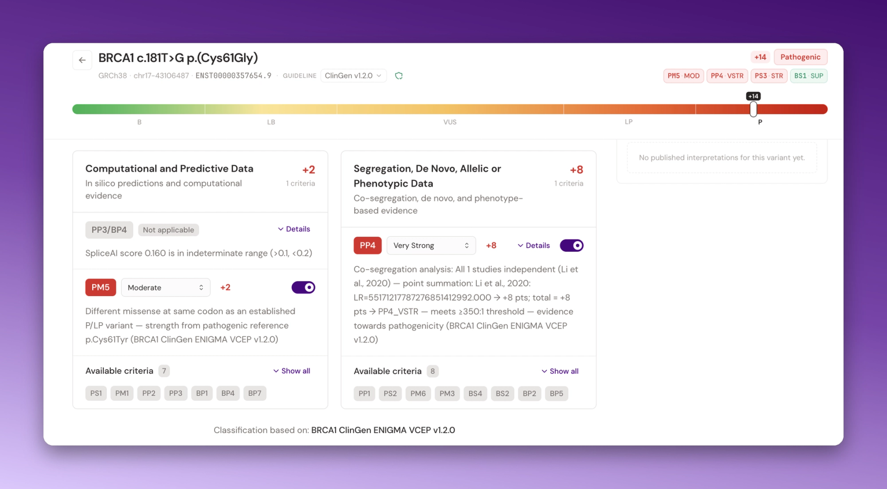
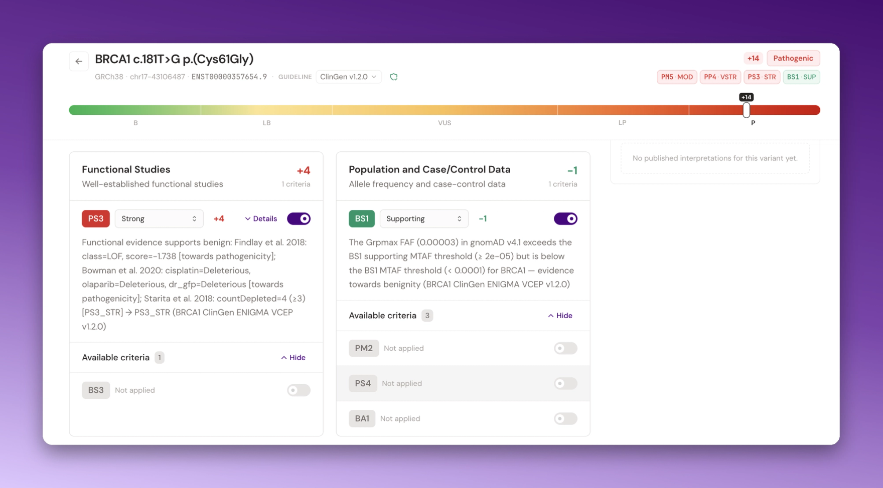
- c.4956G>A: common enough (2.85% of people in gnomAD) to be benign under the rules on frequency alone. The lab result agrees (Figure 13).
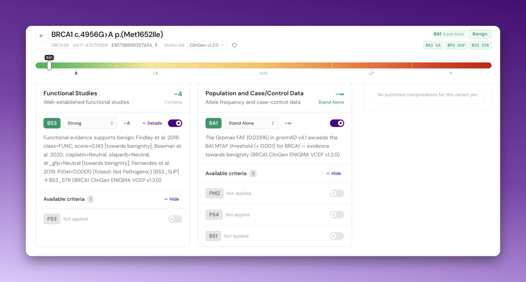
In every case VAIC applies the guideline and lays out the evidence behind each criterion. The clinical team reviews it and makes the call.
Same disease, different biology, and who the data leaves out
Zoom out from one gene to a thousand tumours. In 2012, The Cancer Genome Atlas reported TP53 mutations in about 80% of basal-like breast tumours, with PIK3CA mutations concentrated in the luminal subtypes [6]. Does that still hold on today's public data?
We opened the TCGA PanCancer Atlas breast cancer dataset, breast cancer study on cBioPortal [7], using the 1,066 tumours in its mutation files, in Pan.bio Notebooks and asked bioMind in plain language. It found the files, wrote the code, and we reviewed and ran it (Figure 14).

Across all 1,066 tumours, TP53 and PIK3CA tie as the most frequently mutated genes, at 32.5% each. Split by molecular subtype (PAM50), they run in opposite directions (Figure 15, Table 3):
Table 3. TP53 and PIK3CA mutation rates by PAM50 subtype, TCGA PanCancer Atlas [7].
Subtype | TP53 mutated | PIK3CA mutated |
|---|---|---|
Basal-like (171 tumours) | 88.9% | 7.0% |
HER2-enriched (78) | 70.5% | 32.1% |
Luminal B (197) | 36.0% | 29.9% |
Luminal A (499) | 10.6% | 47.5% |
The 2012 finding holds. Same disease, different biology.

The second question took one more prompt: who is in this data? Of the 1,066 tumours, 795 (74.6%) come from people of European ancestry. Another 150 carry other ancestry labels, 121 have none, and Middle Eastern is not a category at all (Figure 16).

Every result above describes the people who were sampled. The same few cells, run on tumours from this region, would show whether these patterns hold here too. The code is ready. The data is what is missing.
Three scales, one place, every step on the record
What the three analyses share is not the biology. It is how they were run: the raw reads, the code, the results and the evidence sit in one workspace, and every step can be opened, checked and run again: the Pan.bio workflows and notebooks for the analysis, VAIC for the interpretation.
What none of them can fix alone is the reference data. The ancestry gap in the TCGA cohort and the population frequencies VAIC reads for every variant both depend on who was sequenced. Pan.bio is building the infrastructure for more sequencing, bioinformatics and analysis capacity here, so that future answers draw on data that reflects everyone.
The same steps work on your own data. Sign up at pan.bio and run your first analysis.
References
- Findlay GM, et al. Accurate classification of BRCA1 variants with saturation genome editing. Nature 2018;562(7725):217–222. PMC6181777
- Bouwman P, et al. Functional categorization of BRCA1 variants of uncertain clinical significance in homologous recombination repair complementation assays. Clin Cancer Res 2020;26(17):4559–4568. doi:10.1158/1078-0432.CCR-20-0255
- Fang LT, et al. Establishing community reference samples, data and call sets for benchmarking cancer mutation detection using whole-genome sequencing. Nat Biotechnol 2021;39(9):1151–1160. doi:10.1038/s41587-021-00993-6. Xiao W, et al. Toward best practice in cancer mutation detection with whole-genome and whole-exome sequencing. Nat Biotechnol 2021;39(9):1141–1150. doi:10.1038/s41587-021-00994-5
- Richards S, et al. Standards and guidelines for the interpretation of sequence variants. Genet Med 2015;17(5):405–424. PubMed 25741868
- Olsson E, et al. Mutation screening of 1,237 cancer genes across six model cell lines of basal-like breast cancer. PLOS ONE 2015;10(12):e0144528. doi:10.1371/journal.pone.0144528
- The Cancer Genome Atlas Network. Comprehensive molecular portraits of human breast tumours. Nature 2012;490(7418):61–70. doi:10.1038/nature11412
- cBioPortal, Breast Invasive Carcinoma (TCGA, PanCancer Atlas). cbioportal.org
- ClinVar: VCV 55480 (c.5251C>T), VCV 17661 (c.181T>G), VCV 41830 (c.4956G>A)
- Parsons MT, et al. Evidence-based recommendations for gene-specific ACMG/AMP variant classification from the ClinGen ENIGMA BRCA1 and BRCA2 Variant Curation Expert Panel. Am J Hum Genet 2024;111(9):2044–2058. doi:10.1016/j.ajhg.2024.07.013. Current specifications: ClinGen CSpec registry

Bioinformatician with 5 years of experience in analyzing biomedical and clinical data. I connect genomics with clinical picture using data science, machine learning, and a purpose-driven mindset.
View full profile
236 Sarcomas Profiled in One Sitting. Two Routes to One Broken Pathway
We ran a full multi-omic sarcoma analysis in a single session of Pan.bio Notebooks. Every step was a plain-English question to bioMind. The most interesting result was one we never asked for.
How to Choose Clinical Bioinformatics Software: 8 Questions to Ask Before You Buy
How to evaluate clinical bioinformatics software before you buy: 8 questions covering validation, AI governance, data residency, population coverage, cost, and vendor lock-in.
Your Genome Was Never in the Study, Only 1 in 600: Why Arab DNA Is the Biggest Blind Spot in Modern Medicine
Arab genetic architecture is its own genomic landscape, shaped by thousands of years of a unique demographic history, geographic isolation, and cultural practices that have no real parallel in the populations that dominate global genomic databases.
Ahmad Jadallah6 min